📄 Publications
Preprints & Under Review
Hierarchical Enzyme Classification with Integrated Sequence and Folding-Derived Representations
Po-Yu Liang, Wei Wang, Jun Bai*
Submitted to ACM BCB 2026
PepEDiff: Out-of-Distribution Sampling Peptide Binder Design via Protein Embedding Diffusion
Po-Yu Liang, Jun Bai*
Submitted to ACM BCB 2026
Uncovering the Mechanism of Hepatotoxicity of PFAS Targeting L-FABP Using GCN and Computational Modeling
Lucas Jividen, Tibo Duran, Xi-Zhi Niu, Jun Bai*
arXiv:2409.10370 · 2024
A Tonic Signaling Code Predicts CAR-T Cell Efficacy in Diffuse Midline Glioma
Emily B Deng, Xiaowen Zhong, ..., Po-Yu Liang, Jun Bai, et al.
bioRxiv · 2025
MultiST: A Cross-Attention-Based Multimodal Model for Spatial Transcriptomics
Wei Wang, Chong Yu, Jun Bai*
bioRxiv · 2026
Refereed Journal Articles
Hybrid Transformer-based Model for Mammogram Classification by Integrating Prior and Current Images
Afsana Ahsan Jeny, Sahand Hamzehei, Annie Jin, Stephen Andrew Baker, Tucker Van Rathe, Jun Bai, Clifford Yang, Sheida Nabavi
Medical Physics · 2025 | IF: 4.506
Advanced Feature Extraction and Outlier Detection for 3D Biological/Biomedical Image Registration
Sahand Hamzehei, Jun Bai, Gianna Raimondi, Rebecca Tripp, Linnaea Ostroff, Sheida Nabavi
IEEE Transactions on Computational Biology and Bioinformatics · 2025 | IF: 3.71
Unsupervised Feature Correlation Model to Predict Breast Abnormal Variation Maps in Longitudinal Mammograms
Jun Bai, Annie Jin, Madison Adams, Clifford Yang, Sheida Nabavi
Computerized Medical Imaging and Graphics · Vol. 113 · 102341 · 2024 | IF: 5.7
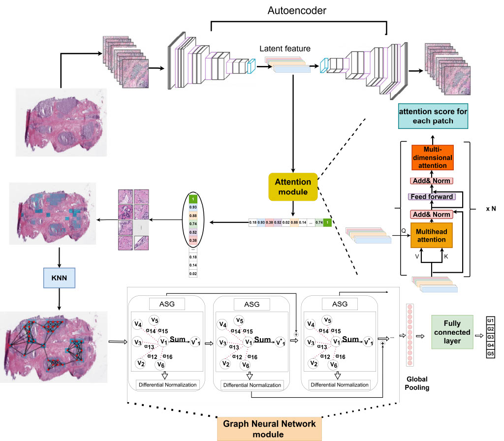
Weakly-Supervised Deep Learning Model for Prostate Cancer Diagnosis and Gleason Grading of Histopathology Images
Mohammad Mahdi Behzadi, Mohammad Madani, Hanzhang Wang, Jun Bai, et al.
Biomedical Signal Processing and Control · Vol. 95 · 106351 · 2024 | IF: 4.9
Molecular Dynamics Modeling Based Investigation of the Effect of Freezing Rate on Lysozyme Stability
Tibo Duran, Bruna Minatovicz, Ryan Bellucci, Jun Bai, Bodhisattwa Chaudhuri
Pharmaceutical Research · Vol. 39(10) · 2585–2596 · 2022 | IF: 4.914
Combining Multi-view Ensemble and Surrogate Lagrangian Relaxation for Real-Time 3D Biomedical Image Segmentation on the Edge
Shanglin Zhou, Xiaowei Xu, Jun Bai, Mikhail Bragin
Neurocomputing · Vol. 512 · 466–481 · 2022 | IF: 5.719
Feature Fusion Siamese Network for Breast Cancer Detection Comparing Current and Prior Mammograms
Jun Bai, Annie Jin, Tianyu Wang, Clifford Yang, Sheida Nabavi
Medical Physics · Vol. 49(6) · 3654–3669 · 2022 | IF: 4.506
Molecular Dynamics Simulation to Uncover the Mechanisms of Protein Instability During Freezing
Tibo Duran, Bruna Minatovicz, Jun Bai, Dongkwan Shin, Hossein Mohammadiarani, Bodhisattwa Chaudhuri
Journal of Pharmaceutical Sciences · Vol. 110(6) · 2457–2471 · 2021 | IF: 4.064
Single-cell Classification Using Graph Convolutional Networks
Tianyu Wang, Jun Bai, Sheida Nabavi
BMC Bioinformatics · Vol. 22(1) · 1–23 · 2021 | IF: 4.341
Applying Deep Learning in Digital Breast Tomosynthesis for Automatic Breast Cancer Detection: A Review
Jun Bai, Russell Posner, Tianyu Wang, Clifford Yang, Sheida Nabavi
Medical Image Analysis · Vol. 71 · 102049 · 2021 | IF: 13.828
Conference Papers
DeepPH: A Multimodal Deep Learning Model for Predicting Enzyme Optimal pH Range
Wei Wang, Po-Yu Liang, Jun Bai*
16th ACM BCB · 2025 | Acceptance rate: ~19%
Robust Training of Deep Learning Models for Mammogram Classification
Josue Martinez-Martinez, Olivia Brown, Jun Bai, Sheida Nabavi
IEEE 22nd ISBI · 2025 | Acceptance rate: ~25%
E(3)-Invariant Diffusion Model for Pocket-Aware Peptide Generation
Po-Yu Liang, Jun Bai*
ISBRA 2025 · Springer LNBI vol. 15757 · pp 177–189 | Acceptance rate: ~19%
Exploring Latent Space for Generating Peptide Analogs Using Protein Language Models
Po-Yu Liang, Xueting Huang, Tibo Duran, Andrew J. Wiemer, Jun Bai*
IEEE BIBM 2024 · pp 842–847 | Acceptance rate: ~19%
3D Biological/Biomedical Image Registration with Enhanced Feature Extraction and Outlier Detection
Sahand Hamzehei, Jun Bai, Gianna Raimondi, Rebecca Tripp, Linnaea Ostroff, Sheida Nabavi
14th ACM BCB · 2023 | Acceptance rate: ~19%
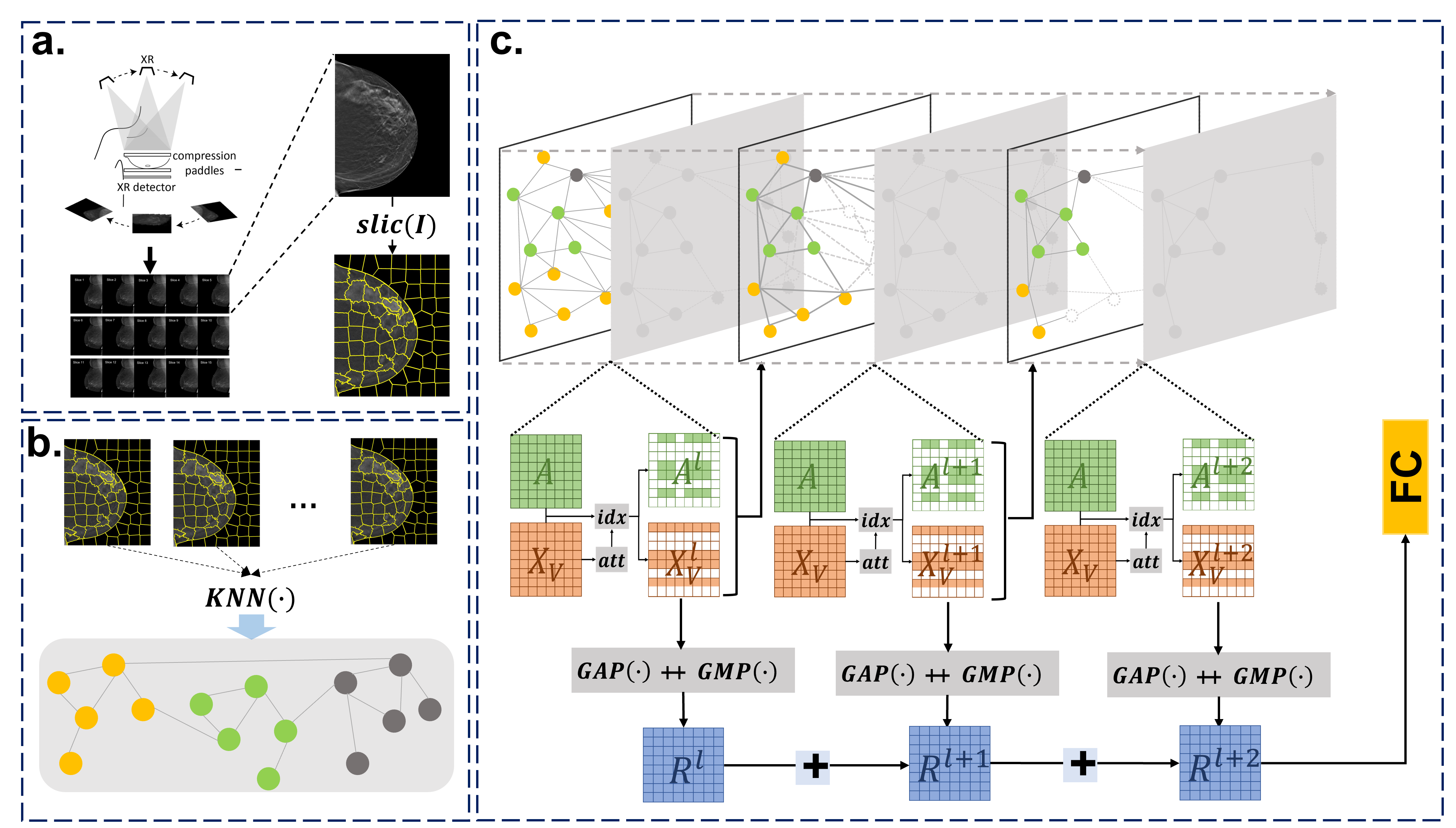
Applying Graph Convolution Neural Network in Digital Breast Tomosynthesis for Cancer Classification
Jun Bai, Annie Jin, Andre Jin, Tianyu Wang, Clifford Yang, Sheida Nabavi
13th ACM BCB · 2022 | Acceptance rate: ~19%
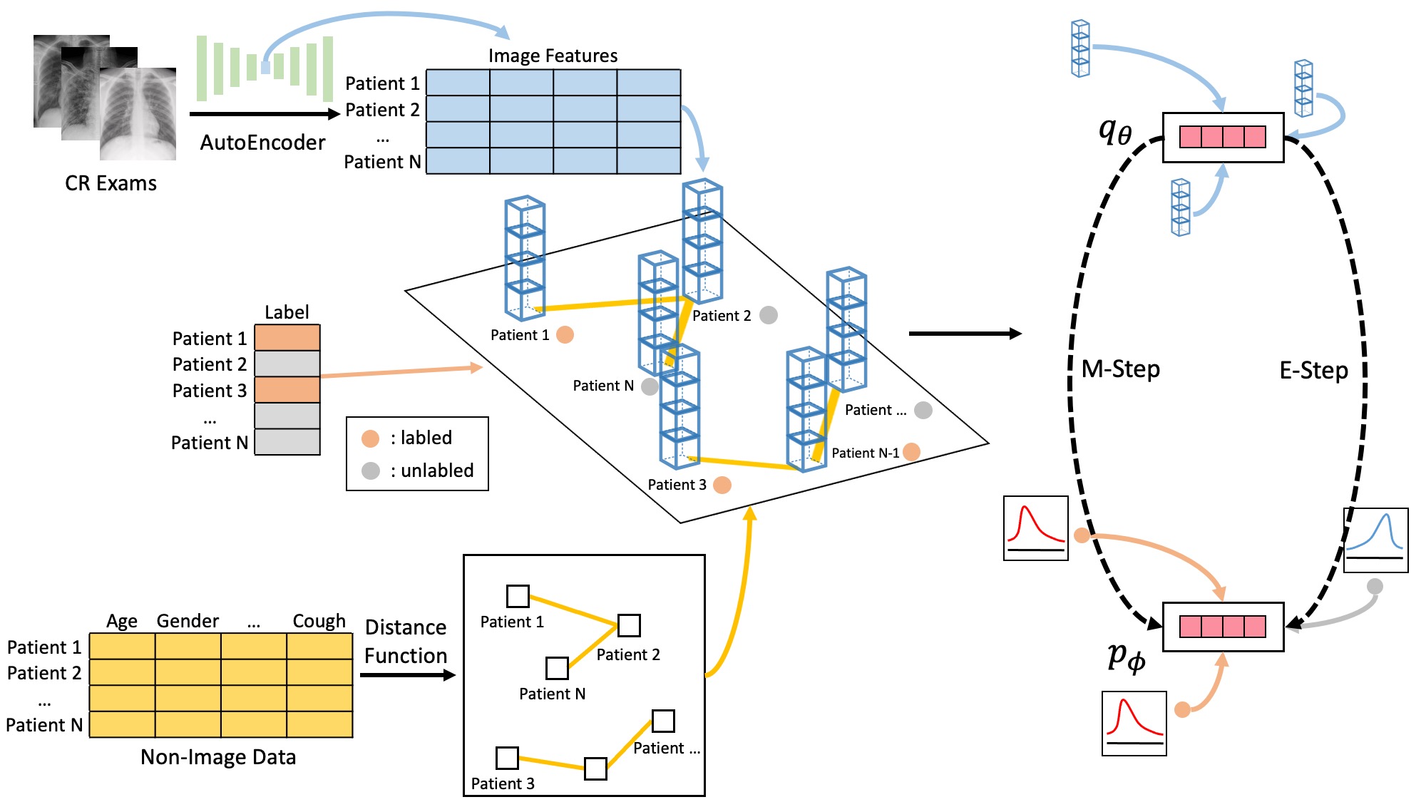
Semi-supervised Classification of Disease Prognosis Using CR Images with Clinical Data Structured Graph
Jun Bai, Bingjun Li, Sheida Nabavi
13th ACM BCB · 2022 | Acceptance rate: ~19%
A Deep Learning Approach for Ventricular Arrhythmias Classification using Microcontroller
Ya-Sine Agrignan, Shanglin Zhou, Jun Bai, Sheida Nabavi, Caiwen Ding
International Symposium on Quality Electronic Design · 2023
Patent
US 12387477 — Conjoined Twin Network for Breast Cancer Treatment and Analysis
United States Patent
Full list: Google Scholar ↗ | ResearchGate ↗